

![]()
Bigelow Laboratory for Ocean Sciences: A.i.R.

Bigelow Laboratory for Ocean Sciences (BLOS) is a nonprofit research institute in East Boothbay, Maine using innovative methodological and technological approaches to study global ocean health. In 2016, I was the initiator and first ever participant of their artist in residence program, pairing visual artists with members of BLOS’ research staff to explore intersections of art, science and communication. For four months I worked alongside senior research scientist Dr. Steve Archer and research associate Carlton Rauschenberg developing a visually immersive and experiential interpretation of Archer's research on ocean acidification and its impact on microbiological production of dimethyl sulfide (DMS). DMS, once aerosolized, is important in the formation of planetary cooling cloud coverage.


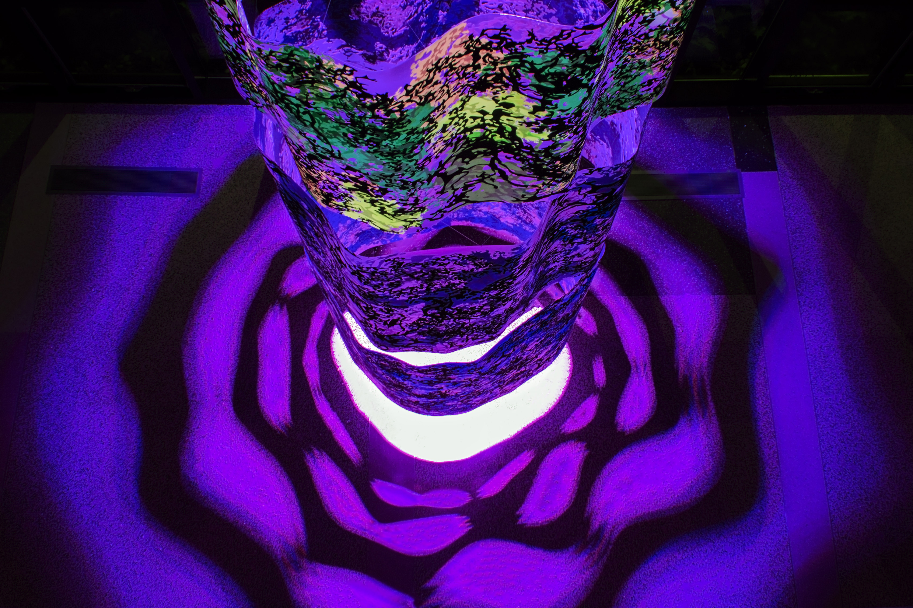
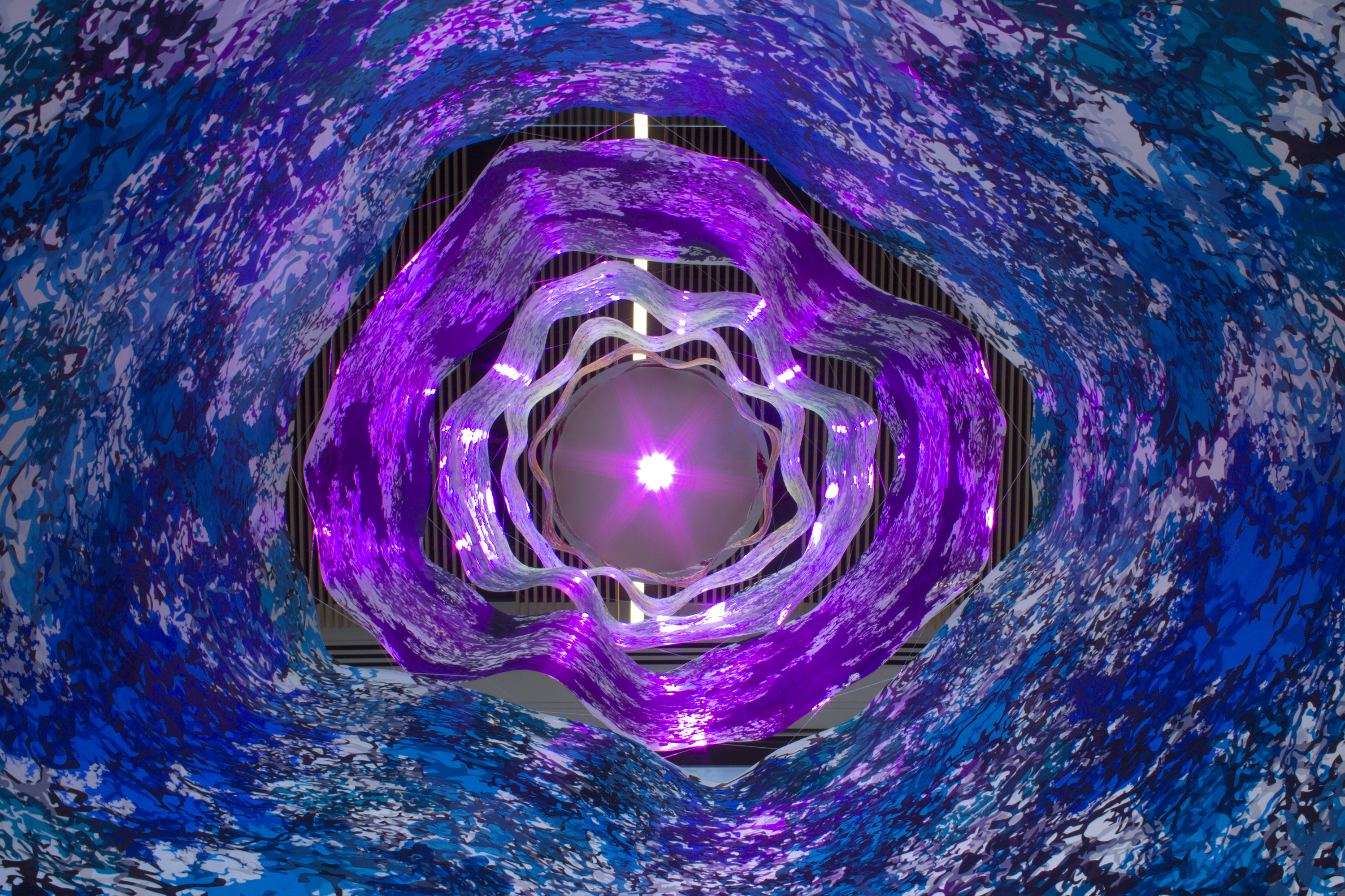
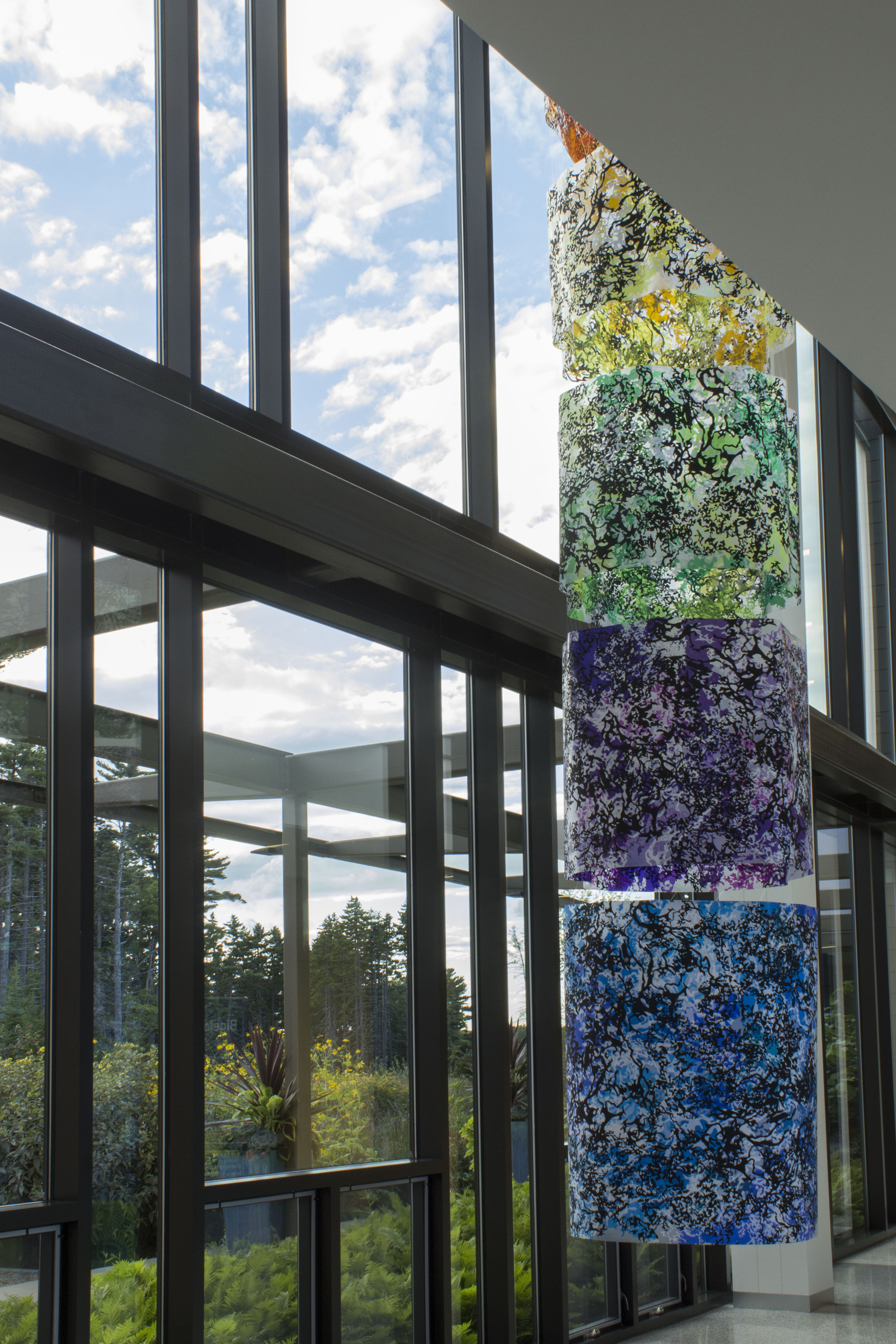

Colorcosm
Screen print on sanded plastic, monofilament, LED light & wooden cap.
4' diameter x 21'
2016
The result of our collaboration is Colorcosm, a 21’ tall screen printed sculpture consisting of six floating, prismatic plastic cylinders. Colorcosm, through visual and conceptual nods, serves as a doorway to the complex information presented in Archer’s research. The sculpture drew inspiration from our expansive dialogue on the nature of art and science, printmaking, visible light, gas exchanges, algal pigmentation, acids, experimental field technology, copper etching and a series of scanning electron microscope images (below). Colorcosm, allows the landscape and architecture of the laboratory to flow through the piece, while offering a new perspective—a kind of fantastic lens to see through. It reminds us of the greater context which the piece is situated in. It pulls us outwards to the water and upwards to the atmosphere. For more information on my time at the laboratory and the creation of Colorcosm visit my blog, which was maintained for the duration of the residency.
SEM Images
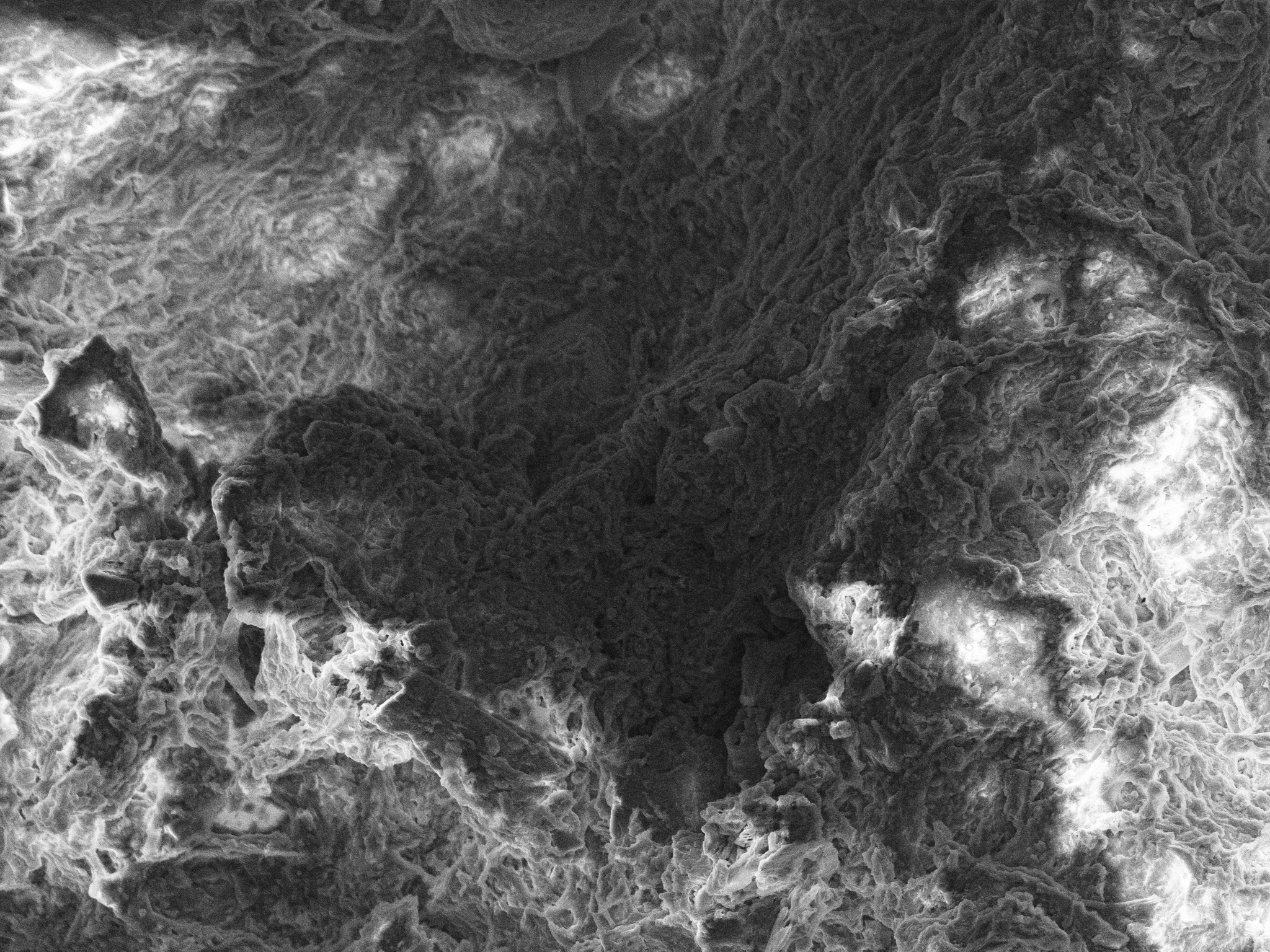
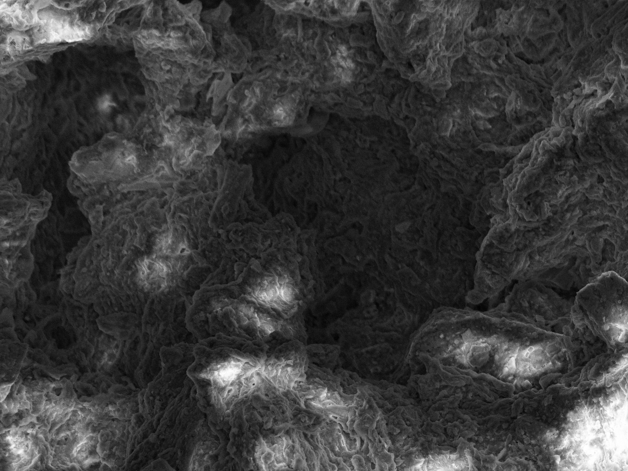
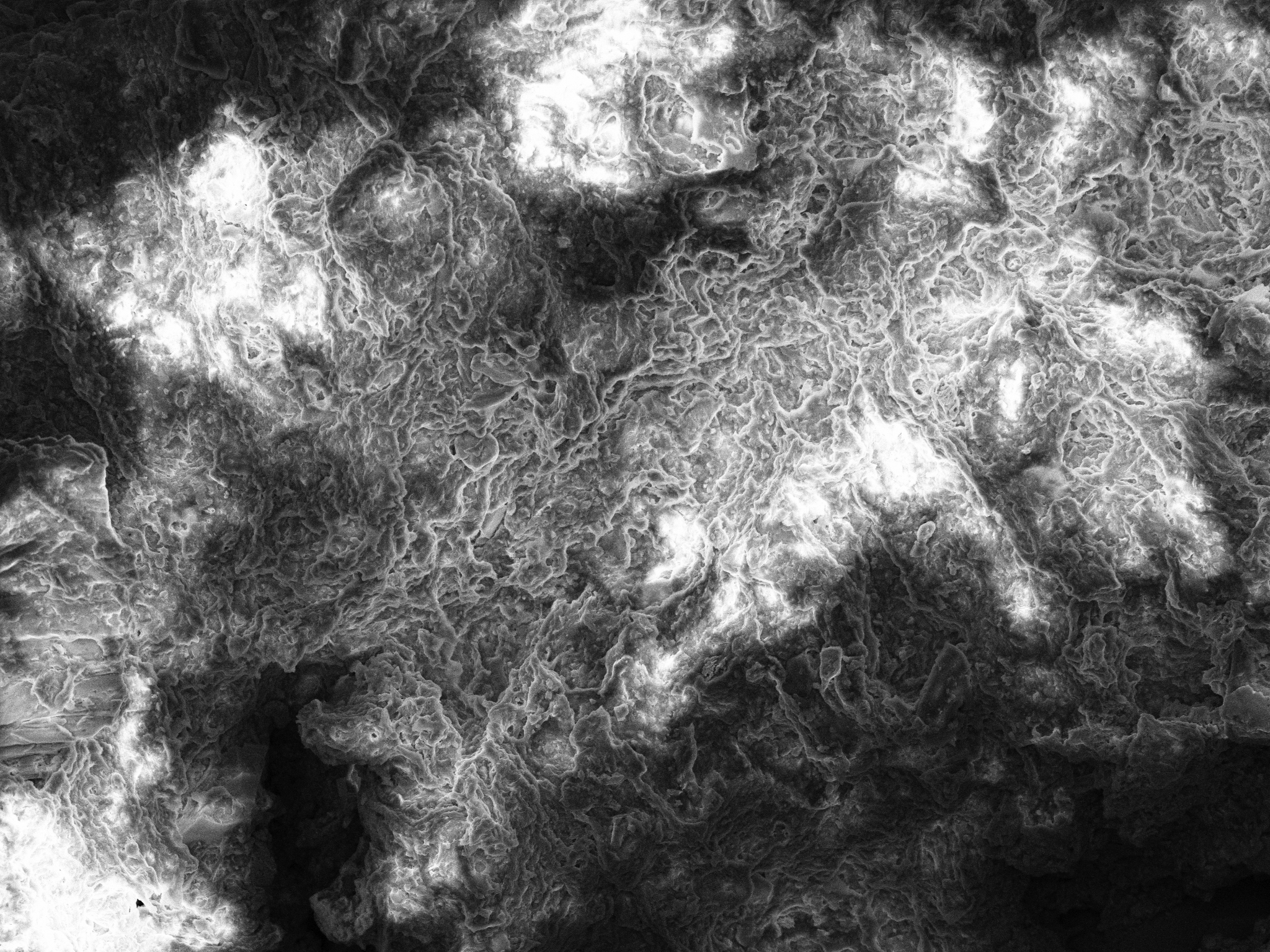
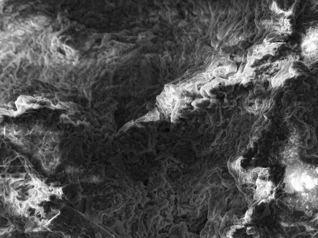
Untitled SEM Images
Scanning electron microscope image
2016
These images were taken using BLOS’ scanning electron microscope with the guidance of research associate Carlton Rauschenberg. The imaged samples included flotsam rope (which I’ve collected over the years for my Ghost Gear series), coralline algae and tiny etched copper fragments. Typically, samples for scanning are lightly sputter coated in gold prior to imaging. The gold coating reflects a beam of electrons, used to collect visual data of the sample, back to the machine’s sensors. An unevenly or insufficiently coated sample will cause some of the reflected electrons to charge-up due to contact with exposed organic matter on the sample. The result of this can be seen throughout the images above in the seemingly artificial light, or, what Rauschenberg termed, "flare-ups.”
For the kinds of precise and consistent imaging necessary for scientific research, these poorly coated samples would need to be resampled or recoated. However, I found the unpredictability of such samples to produce images with greater depth and spatiality. An almost theatrical or cinematic effect occurs, where the lines of photography, scientific imaging, fantasy and reality are blurred.
For the kinds of precise and consistent imaging necessary for scientific research, these poorly coated samples would need to be resampled or recoated. However, I found the unpredictability of such samples to produce images with greater depth and spatiality. An almost theatrical or cinematic effect occurs, where the lines of photography, scientific imaging, fantasy and reality are blurred.